Elbow Instability – Collateral ligament injury
The notes on this page are written as a source of information to both patients and health professionals. It is a general overview and its content should not be seen as direct medical advice. Further information regarding diagnosis and treatment specific to your condition can be made via a consultation with Dr Amaranath.
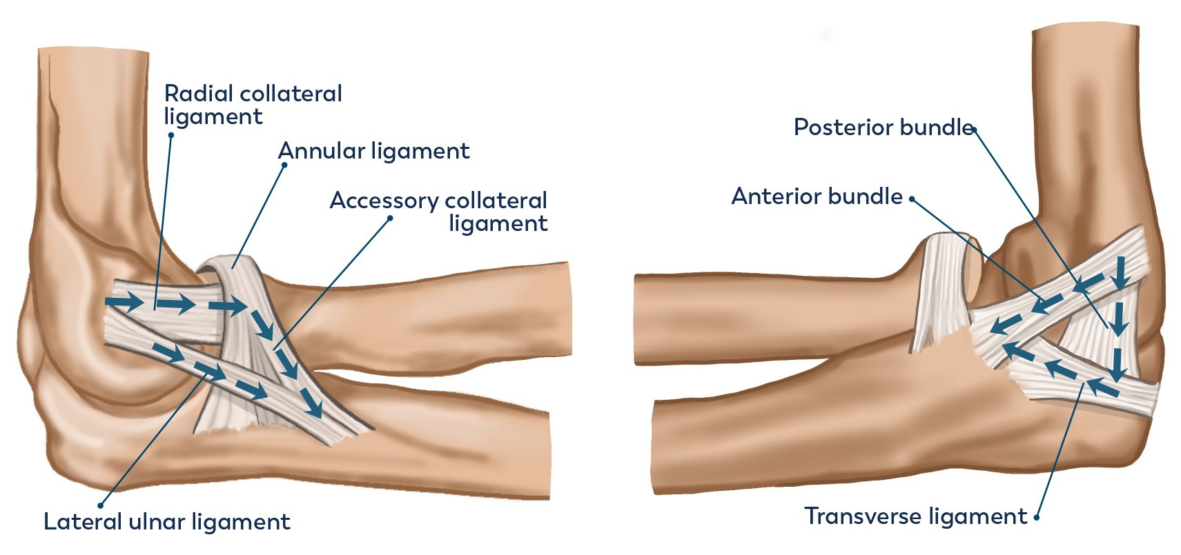
Anatomy of the elbow
The elbow acts as a hinge joint allowing movements of flexion and extension. The joint is kept stable by the bony articulation between the olecranon and the trochlea, along with the medial and lateral collateral ligaments. The lateral ligaments protect against varus instability (moving towards the body) and are made of 4 primary ligaments. The Lateral Ulna Collateral Ligament (LUCL) is the most important. The Medial Collateral Ligament (MCL) protects against valgus instability and is made of 3 components, of which the anterior bundle is the most significant.
What is elbow instability?
This refers to injury to one or both of the collateral ligaments leading to asymmetrical motion within the elbow joint. In some patients this can manifest as pain or apprehension (fear) in certain movements depending on the ligament that is injured.
What are the common causes?
Trauma – Injury to the LUCL or MCL is commonly seen in elbow dislocations following a traumatic event e.g. fall on outstretched hand, wrestling, etc.
Overuse – This is due to repetitive stress of the ligament due to certain activities e.g. throwing, bowling, etc.
What are the preferred Investigations/Imaging?
Elbow X-ray – This should be the first imaging modality used as it helps determine if there is any underlying arthritis, associated fractures or other pathology that may be a cause of the symptoms.
MRI – This is the best form of imaging to identify a collateral ligament injury and can also asses joint congruity.
What are the treatment options?
Non-operative – This depends on the degree and type of injury. In strains of the collateral ligament, non-operative measures such as a graduate physiotherapy programme, analgesia and activity modifications can be utilised.
Elbow stabilisation – Surgery is indicated in a traumatic disruption of the ligament, failed non-operative management or recurrent instability limiting the patient’s function. It involves an open surgical procedure where the ligament in anchored back onto the distal humerus (origin) or ulna (insertion) through the use of anchors and suture material. If the injury is chronic then a reconstruction is needed (as the old ligament is unlikely to be able to be reattached), which utilises a tendon graft as a substitute.
What does the rehabilitation/recovery involve?
To find out more about rehabilitation and recovery after elbow surgery please see our Rehabilitation Protocols here.